
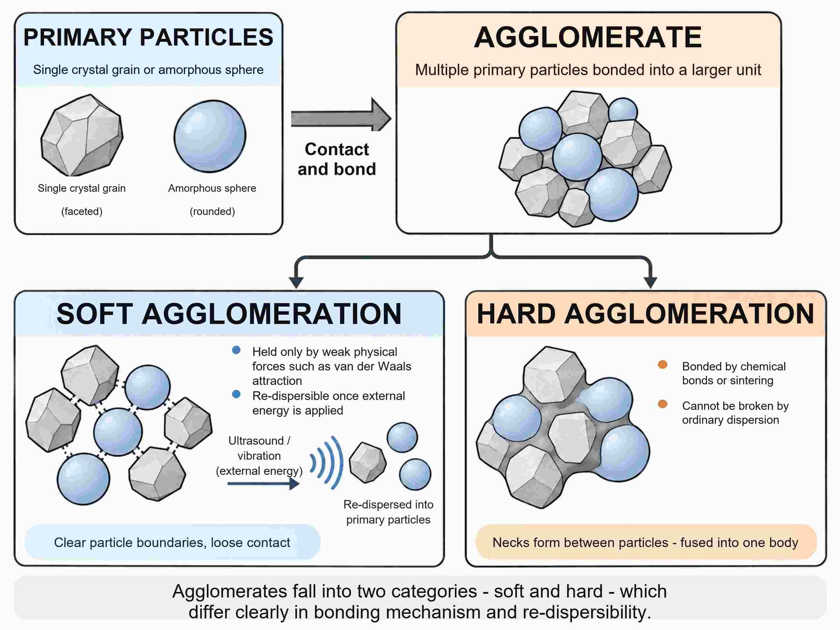
Synthesis produces single crystal grains or amorphous spheres, each of them a primary particle. Let several of them touch and bond, and they form a larger unit — an agglomerate.
Agglomerates differ in how firmly they are held together. In a soft agglomerate the primary particles are bound mainly by weak physical forces such as van der Waals attraction, and ultrasound or another source of external energy can separate them again. In a hard agglomerate the contact points have been welded shut by chemical bonds or by sintering, and no ordinary dispersion step will prise them apart.
Size drives the whole process. The finer the particles, the greater the share of atoms sitting at the surface, and the greater the specific surface area and the surface free energy of the system. Whenever two particles touch, part of that surface is replaced by a contact interface and the free energy falls. Agglomeration is thus the energetically favourable outcome.
Commercial γ-Al₂O₃ nanopowders behave in exactly this way. Powders sold as 5 nm and 10 nm material both showed primary particles of about 10 nm under the TEM, and even after 30 min of probe sonication in 1 mM NaCl the hydrodynamic diameter in acid remained at 202–266 nm.
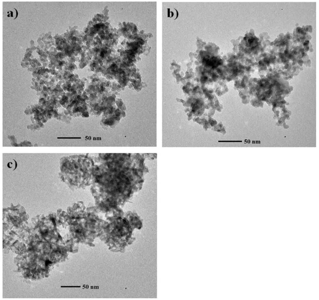
Figure 1. Bright-field TEM images of three commercial γ-Al₂O₃ nanoparticle samples (nominal sizes 5 nm, 10 nm and 20–30 nm). DOI: 10.3390/nano6050090
The gap between the two numbers comes from what each technique actually measures. TEM reports the projected size of a single primary particle. Dynamic light scattering follows the way the intensity of the scattered light fluctuates in time and converts that, through the Stokes–Einstein relation, into a hydrodynamic diameter — the size of the object that really moves through the liquid.
Because the γ-Al₂O₃ hydrodynamic diameter is more than twenty times the primary particle size, every moving unit must contain many primary particles. Not all of the clumping seen in a micrograph is pre-existing, however: some of the contacts are created on the grid as the droplet dries.
Those new contacts come from solvent evaporation. As the liquid retreats, bridges form between the particles and shrink, and the resulting capillary force is strong enough to override the electrostatic repulsion that had kept them apart. In the liquid itself the balance is different, being set by van der Waals attraction competing with electric double layer repulsion and with steric hindrance from adsorbed layers. Heat the powder to calcination or reaction temperature and the picture changes once more: sintering necks grow at the contact points, atoms diffuse across the interface, and neighbouring particles merge into one larger particle.
Bring two nanoparticles together in a liquid and the van der Waals attraction climbs steeply as the gap narrows. Classical DLVO theory adds that attraction to the electric double layer repulsion between the particles; if the repulsion is strong enough, the sum of the two forms an energy barrier that most encounters never get past. Two quite different mechanisms can supply such repulsion — surface charge, which acts electrostatically, and adsorbed polymer, which acts sterically — and they respond to added salt in different ways.
Oxide particles in water carry hydroxyl groups on their surface. Protonate them at low pH and the particles turn positive; strip the protons off at high pH and they turn negative. The pH at which the zeta potential passes through zero is the isoelectric point.
For titanium dioxide P25 that point lies at about pH 6.2, and it does not move when the NaCl concentration is changed, because Na⁺ and Cl⁻ do not specifically adsorb on the surface. Near pH 6.2 the net surface charge is vanishingly small and large flocs appear in the dispersion.
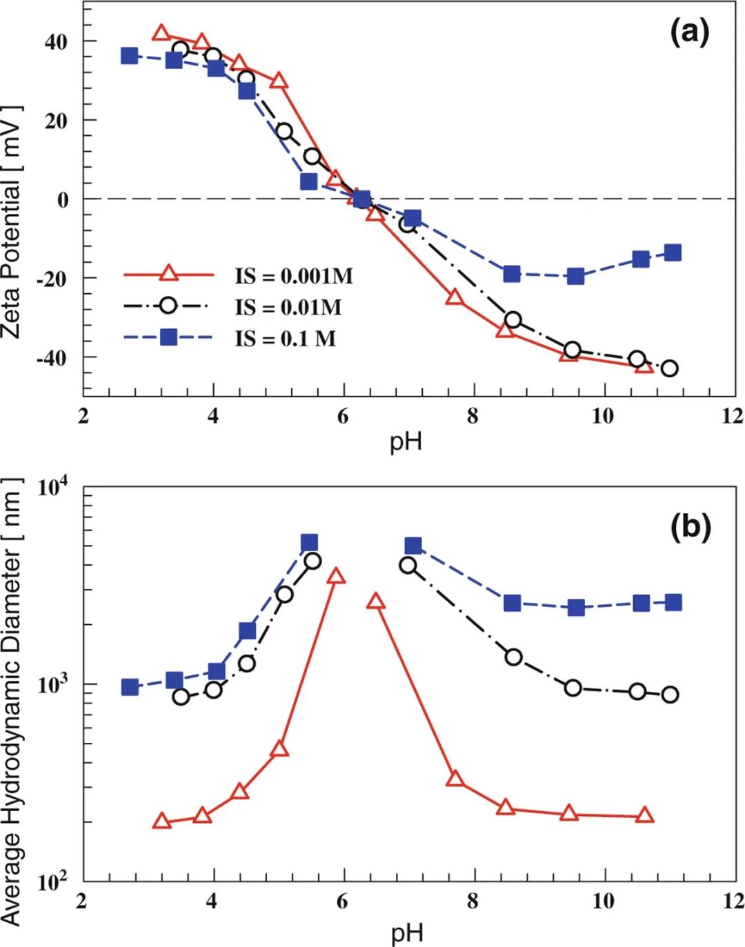
Figure 2. Zeta potential and average hydrodynamic diameter of titanium dioxide nanoparticle dispersions as a function of pH in NaCl solutions of different ionic strengths. DOI: 10.1007/s11671-010-9772-1
Salt acts by compressing the double layer around each particle. Away from the isoelectric point, more ionic strength means a lower absolute zeta potential and a stronger tendency to aggregate. At 0.001 M, P25 dispersions measure roughly 200 nm at pH 3–4 and above pH 8.5; raise the ionic strength to 0.01 M or 0.1 M and the pH 3–4 figure climbs to about 1 μm.
How sensitive a system is to salt depends on the material. γ-Al₂O₃ below its point of zero charge, around pH 7.5–8, tolerates NaCl up to 100 mM without change but aggregates noticeably at 1000 mM.
An electrostatically stabilized dispersion can therefore be protected in two ways: shift the pH away from the isoelectric point, and wash out the salts left over from synthesis with deionized water. Neither helps when the medium is itself salty. At pH 7.4 in 150 mM NaCl — physiological salinity — citrate-stabilized gold nanoparticles aggregate, because part of their surface charge is neutralized.
Polymers offer a second line of defence that owes nothing to electrostatics. Chains firmly anchored on the particle surface and dangling into the solvent force two approaching particles to compress each other's adsorbed layers, and that costs energy. The result is steric repulsion, and it still works once the surface charge has been screened.
The experiment is striking. A dispersion of 15 nm gold nanoparticles was brought to pH 7.4 with 150 mM NaCl. Samples with no PEG, and samples with linear PEG carrying hydroxyl or methoxy end groups, turned from red to grey-blue within moments and settled out; samples with thiol-terminated linear PEG or with cyclic PEG were still red after 1000 min. The authors' explanation is that the adsorbed PEG layer keeps the particles apart even after the salt has screened their charge.
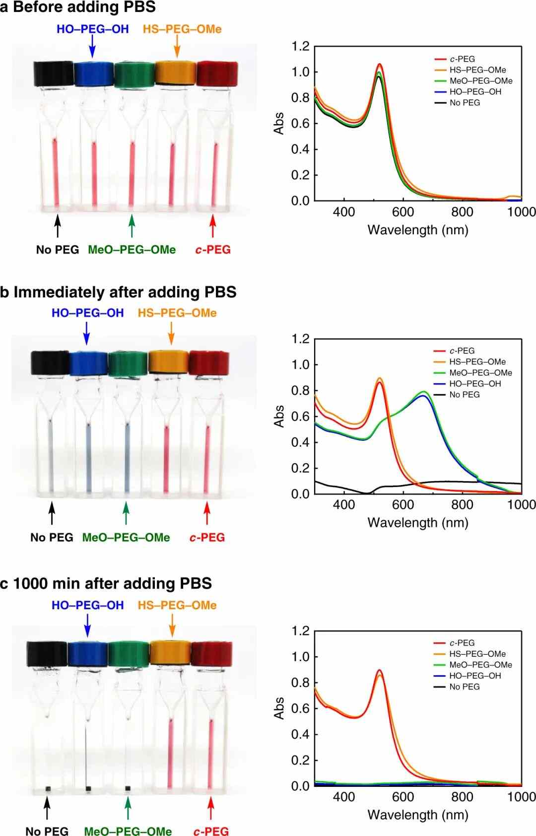
Figure 3. Photographs and UV–vis absorption spectra of gold nanoparticle dispersions containing different polyethylene glycols, before adding concentrated phosphate buffer, immediately after adding it, and after 1000 min. DOI: 10.1038/s41467-020-19947-8
How thick is that layer? Dynamic light scattering gives an estimate. Bare gold particles measured 19 nm across; adding linear PEG with hydroxyl or methoxy ends moved the figure only to 20 nm. Thiol PEG, which binds chemically to gold, took it to 28 nm, and cyclic PEG, held only by physisorption, to 26 nm — evidence that these two build a substantially thicker shell around each particle.
Small molecules fail on the same principle. 18-crown-6 is only about 0.7 nm across, and adding it precipitated every particle.
So does the amount of polymer. With 5 nm gold particles and cyclic PEG of molecular weight around 5000, a concentration of 0.025 wt% left just 22% of the relative absorbance after 1000 min at physiological salinity; 0.25 wt% left 78%, and 2.5 wt% left the value essentially unchanged. Too little coverage can also bridge particles instead of separating them.
The same trap appears during drying. In drop-drying experiments with gold particles, using bovine serum albumin at about one twentieth of the optimal dose allowed individual protein molecules to adsorb on several particles at once, knitting them into large clusters.
Solvent removal is where soft agglomerates can turn into hard ones. As the powder dries, the liquid retreats into the gaps and forms bridges whose curved surfaces pull the particles together.
The forces involved are not small. For two 20 nm silica particles the liquid bridge energy during evaporation has been estimated at about 10³ kBT — far above a typical electrostatic barrier, and enough to pull the particles into the range where van der Waals attraction takes over. As the last of the water leaves the surface, the attraction strengthens further.
The Young–Laplace equation, ΔP = 2γcosθ/r, sets the scale of the problem: γ is the surface tension of the solvent, θ the contact angle and r the pore radius. Small pores mean large capillary pressures, and the gaps between nanoparticles are measured in nanometres.
Two levers follow from the equation. One is γ: exchanging the water between the particles for ethanol or another low-surface-tension solvent before drying lowers the capillary pressure. The other is θ: silica wet gels, whose surface carries silanol groups, are often treated with a silylating reagent such as trimethylchlorosilane before ambient-pressure drying. The reagent swaps the hydrogen of Si–OH for alkyl groups such as methyl, making the surface hydrophobic, raising the contact angle and reducing the pressure again.
Capillary pressure is only part of the story. As a bridge shrinks, small particles migrate towards the contact point and are deposited there; in a polydisperse silica system spanning 20 nm to 90 μm they form solid bridges between the large particles once the powder is dry. In bidisperse and polydisperse deposits the detachment energy of a 20 μm particle is roughly 15 and 20 times that in a monodisperse system, and the clusters resist both re-wetting and scouring by a water stream.
Both the capillary force and the solid bridges originate at the retreating liquid surface, so the way to avoid them is to do away with that surface. Supercritical drying does exactly that: temperature and pressure are taken above the critical point of the solvent, the liquid becomes a supercritical fluid, and with the liquid–gas interface gone the surface tension — and the capillary force with it — disappears. In low-temperature supercritical drying the water is first exchanged for an organic solvent such as ethanol and then for CO₂, whose critical temperature is near room temperature; the price is pressure-resistant equipment and higher cost.
Freeze-drying takes the other route, freezing the water and then subliming it at about one ten-thousandth of atmospheric pressure, so that liquid water never evaporates. Freezing, however, creates difficulties of its own: as ice forms, solutes concentrate in the shrinking volume of unfrozen liquid, interparticle interactions intensify, and the growing ice crystals impose mechanical stress.
For 15 nm gold nanoparticles frozen in liquid nitrogen and freeze-dried, the outcome mirrors the earlier salt experiment. Samples with no PEG, or with hydroxyl- or methoxy-terminated linear PEG, came out as grey-blue solids that would not re-disperse in water, their relative absorbance no higher than 9%. The thiol PEG and cyclic PEG samples stayed red and recovered to 84% and 91% respectively once re-dispersed.
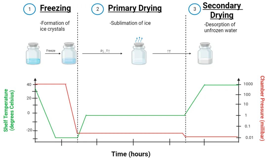
Figure 4. State of the samples in the three stages of freeze-drying — freezing, primary drying and secondary drying — and schematic of the shelf temperature and chamber pressure as a function of time. DOI: 10.3390/ijms241814041
In practice, PEG is less often the answer than a lyoprotectant such as trehalose or sucrose, thought to form a glassy matrix around the particles at low temperature — keeping them apart and cushioning the stress of ice-crystal growth. Primary drying matters too: if the product temperature rises above the collapse temperature, the porous cake collapses, residual moisture climbs and re-dispersion becomes harder.
Ultrasound is the usual way to reopen soft agglomerates in a dried powder, and the dose is measured as energy per unit volume (J/mL). One study of a titanium dioxide pigment dispersed the sample at 562 J/mL in water containing 2% polyphosphate; even so, agglomerates reappeared after standing overnight. Treating it for 1 min in an 80 W ultrasonic bath before measurement left a larger share of primary particles in the size distribution than shaking by hand alone.
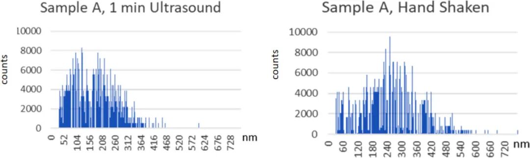
Figure 5. Particle size distributions measured by single-particle inductively coupled plasma mass spectrometry for a titanium dioxide pigment pre-dispersion after 1 min of ultrasonic bath treatment and after re-dispersion by hand shaking only. DOI: 10.3762/bjnano.15.29
Why 562 J/mL? Because above roughly 250 J/mL hard agglomerates and primary particles do begin to break up, but slowly, and further energy may fracture primary particles themselves, biasing the measured size downwards. Some hard agglomerates survive even a thorough dispersion.
Calcination speeds up atomic diffusion, so particles that touch are joined by sintering necks and gradually fuse into larger ones. Supported metal particles have two further routes to growth: an entire particle migrates and merges with a neighbour, or atoms move from a small particle to a large one — Ostwald ripening. Both require the particles to come into contact or to exchange matter.
Separating Particles with a Sacrificial Coating
Consider YVO₄:Yb,Er upconversion nanoparticles made by sodium citrate complexation. They are under 10 nm across but poorly crystalline, so they need annealing — and calcining them directly at 750 °C for 2 h broadens the size distribution and agglomerates them.
A coating solves the problem. A silica shell grown with tetraethyl orthosilicate was in place during the 750 °C, 2 h calcination; soaking in 0.5 M NaOH for 4 h then dissolved most of it away, leaving well-dispersed, highly crystalline particles of 10–15 nm by TEM.
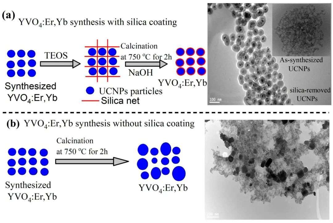
Figure 6. Process of coating YVO₄:Er,Yb upconversion nanoparticles with silica, calcining them and then removing the coating layer with alkali, together with TEM images of the particles obtained by calcination at 750 °C for 2 h with and without the coating. DOI: 10.3390/nano11061535
The mechanism is purely geometric: the shell occupies the space between the particles, so neighbouring YVO₄ particles never touch directly. Crystallinity inside each particle improves, while the contact points needed for sintering necks never form. The NaOH concentration and the soaking time set how much silica is left behind.
For supported metals the question becomes one of distance. Compare d, the distance neighbouring particles must travel to meet, with r, the migration length of a single particle at that temperature. If d < 2r, neighbours meet easily and grow. Geometry then decides how far they can travel: on a flat surface the motion is two-dimensional, while inside a one-dimensional pore it is confined to the pore — although particles sharing a single pore can still meet.
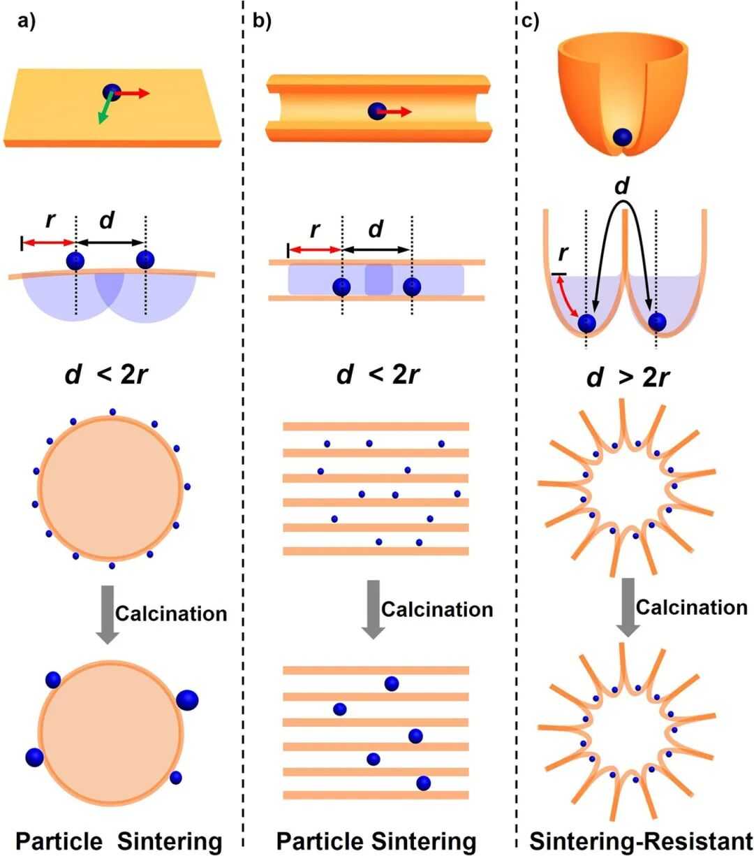
Figure 7. Relationship between the meeting distance d of neighbouring particles and the migration length r of a single particle on three carriers — a flat surface, a one-dimensional pore and a wide-mouth compartment — and schematic of the state of the particles after calcination. DOI: 10.1038/srep41773
Compartmentalized supports exploit this. On the wide-mouth compartment-type silica used here, compartments outnumber platinum particles by more than seven to one, and most hold at most a single particle, so the walls make the true travel distance far longer than the straight-line separation of about 30 nm would suggest.
The numbers bear it out. Platinum nanoparticles of about 1.7 nm, encapsulated in dendrimers and loaded at about 0.07 wt%, were calcined in air at 550 °C for 4 h. On the compartment carrier they were still 1.7 ± 0.4 nm; in the mesopores of SBA-15 they had grown to 7.1 ± 1.8 nm, and on the surface of solid silica nanospheres to 10.1 ± 3.6 nm. Under in-situ heating in vacuum, one particle in a given SBA-15 pore shrank while its neighbour grew — the signature of Ostwald ripening.
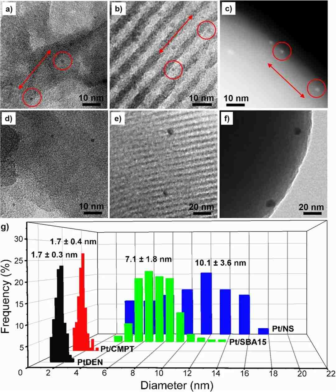
Figure 8. Electron micrographs and particle size distributions of platinum particles supported on compartment-type silica, SBA-15 mesoporous silica and silica nanospheres before and after calcination in air at 550 °C for 4 h. DOI: 10.1038/srep41773
Compartmentalization has limits, and not only for stability. At 0.84 wt% platinum, several particles began to share compartments and grew to 2.7 ± 2.1 nm on calcination. Access, by contrast, improves: CO chemisorption gave platinum dispersions of 67%, 22% and 18% on the compartment carrier, SBA-15 and silica nanospheres respectively. In SBA-15 the platinum also plugs the pores, which lowers the CO oxidation rate once it is normalized to the number of active sites.